
Bright Field vs Phase Contrast vs Fluorescence: Microscope Choice for Biotech Labs
Short answer: choose bright field when the specimen already carries visible contrast — stained tissue, smears, routine morphology. Choose phase contrast when the specimen is alive, unstained and transparent, and contrast has to come from refractive index rather than dye. Choose fluorescence when the biological question is about a specific labelled molecule rather than structure.
Geometry is the second decision. Upright instruments such as the Chongqing Scope B301 and BK5000-PH read specimens on slides, while the inverted BDS500 reads cells through the plastic base of flasks and dishes — the only practical option for most live cell culture work.

Problem Definition: Why Labs Buy the Wrong Contrast Mode
Most microscopy purchasing errors in biotech and medical laboratories are not caused by a bad instrument. They are caused by choosing a contrast mode before the sample state and the specimen container have been defined.
Three recurring mismatches account for most of the cost:
- Fluorescence bought for morphology work. If the question is "what do these cells look like," a filter-based fluorescence platform adds cost and workflow complexity that the study does not require.
- Bright field applied to unstained live cells. Transparent cells in buffer or medium produce very weak contrast in plain transmitted light, so operators compensate with staining — which may change or end the experiment.
- Upright geometry applied to vessels. An upright objective cannot focus through the base of a T-flask or a multi-well plate. No amount of contrast configuration fixes that.
The practical rule is therefore to define four inputs before comparing models: whether the sample is live or fixed, what container it sits in, which contrast mechanism the question requires, and whether the result must be documented digitally.
Industry Background: Optical Contrast Still Sets the Lab Standard
Optical instruments remain the working backbone of life science imaging. According to Market Research Future's 2025 estimate, the global microscope market was valued at approximately USD 11.9 billion, with optical microscopes accounting for roughly 37% of that share. OEC trade data for HS 9011 put global microscope trade at USD 3.36 billion in 2024, with China the third-largest exporter at USD 430 million — a reminder that most biological microscopy hardware in circulation is optical, not electron-based.
Two technical shifts matter for buyers evaluating contrast modes now. First, infinity-corrected optical systems have become the default platform in mid-range laboratory instruments, because they allow filters, condensers and camera adapters to be inserted into the light path without changing the objective's focal behaviour. Second, illumination has moved toward LED sources, which removes the warm-up and lamp-life planning that halogen and mercury systems require.
Both shifts have a procurement consequence: the accessory ecosystem — phase annuli, filter cubes, camera ports — is what actually determines whether one instrument can carry a lab through several workflows, and that ecosystem is model-specific.
Detailed Solution: Matching Documented Models to Contrast Modes
Chongqing Scope Instrument Co., Ltd. is a supplier of laboratory and medical training instruments and metallographic equipment based in Chongqing, China, offering biological, fluorescence, inverted, metallurgical, polarizing and stereo microscopes together with OEM and ODM services. The five models compared below cover the bright field, phase contrast, fluorescence and inverted decision paths.
Bright field: B301 and the infinity-corrected B302 reference
Bright field is the baseline transmitted-light mode: the specimen absorbs or scatters light, and stained structures appear dark against a bright background. It is the correct choice for fixed, stained specimens — H&E sections, blood smears, Gram stains, and routine colony or cell morphology checks — and it is the mode most teaching and screening laboratories standardise on.
The B301 is Chongqing Scope's bright field biological microscope. Its documented compliance position is the notable purchasing fact: the model is covered by CE certificate BKC22072376GC, with the wider product family covered by BKC22072375GC, referenced against EN 61010-1:2010+A1:2019. For a laboratory that needs an EU-market safety file, that is a verifiable document trail rather than a marketing claim.
Where a lab wants a documented infinity optical system in the same bright field role, the B302optical microscope specifies a binocular head, WF10X/20mm eyepieces, infinity achromatic EPlan objectives at 4X/10X/40X/100X, a quadplex nosepiece, a 145×140 mm mechanical stage, an N.A.1.25 Abbe condenser and 3W LED illumination. That specification set is the practical minimum for a general biology bench: four magnifications, a movable stage with measurement reference, and Köhler-style condenser control.
Phase contrast: BK5000-PH for unstained live material
Phase contrast converts differences in refractive index into brightness differences, so transparent specimens become visible without staining. That makes it the default contrast mode for live cells, wet mounts, and any workflow where dye would alter the biology.
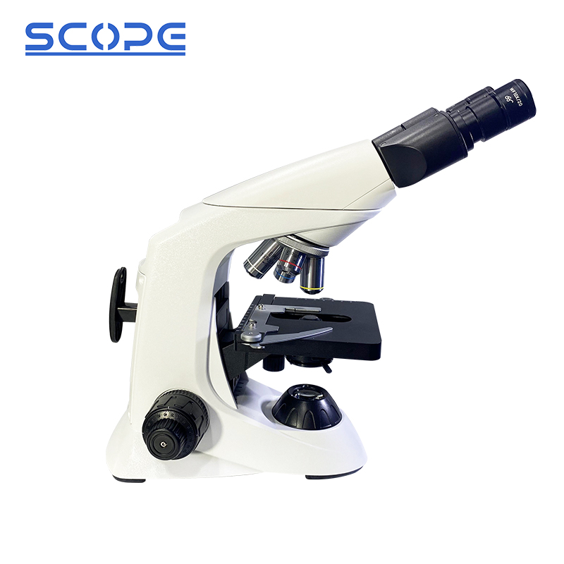
The BK5000-PHphase contrast microscope is specified with a binocular head, WF10X/20mm eyepieces, infinity phase contrast Plan objectives at 4X/10X/40X/100X, a quintuple nosepiece, a 175×145 mm mechanical stage, a quintuple-hole turret Abbe condenser, coaxial coarse and fine focusing, and either 3W LED or 12V/20W halogen illumination.
Two specification details decide whether phase contrast works in practice. The condenser turret must carry a matching annulus for each objective — hence the quintuple-hole turret here — and the annulus must be centred against the objective's phase ring. A lab evaluating this instrument should test centring at 10X and 20X first, because those are the magnifications used for confluency checks and general live-cell inspection. The 100X oil objective supports fine intracellular detail, but it also demands a wet mount with a coverslip, which is not practical inside a closed flask.
The known limitation of phase contrast is optical rather than mechanical: the halo artefact around specimen edges reduces photometric accuracy and can obscure fine detail in thick or highly refractile samples. Labs doing quantitative intensity work should treat phase contrast as a viewing mode, not a measurement mode.
Fluorescence: BK5000-FL for labelled targets
Fluorescence microscopy does not image structure by absorption. It excites a fluorophore with a specific wavelength band and collects the longer-wavelength emission, so only labelled structures appear against a dark background. This is the correct mode when the question is molecular — whether a protein is expressed, where it localises, whether two markers co-localise.
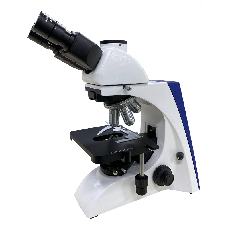
The BK5000-FL fluorescence microscope is specified with a binocular or trinocular head, WF10X/20mm eyepieces, infinity Plan objectives at 4X/10X/20X/40X/100X, a quintuple nosepiece, a 175×145 mm mechanical stage, an Abbe N.A.1.25 condenser with iris diaphragm, and either 3W LED or 12V/20W halogen transmitted illumination. The fluorescence attachment provides blue, green, violet and ultraviolet filters, powered by either a 5W LED or a 100W mercury lamp.
Three specification decisions matter more than the headline "fluorescence" label. The head type determines whether the lab can attach a camera at all: a trinocular head is required for routine documentation, while a binocular head limits imaging to eyepiece-mounted adapters. The filter set must match the fluorophores actually used — a four-channel blue/green/violet/UV set covers common blue, green and UV-excited stains, but a lab working with a narrow far-red dye would need to confirm filter availability before ordering. The lamp choice sets the operating model: LED avoids mercury lamp warm-up, intensity decay and disposal obligations, while a 100W mercury source remains the reference for demanding UV channels.

Inverted geometry: BDS500 and BDS400 for vessel-based work
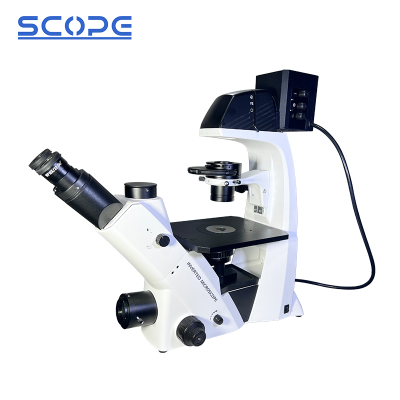
The single most important specifier for cell culture is not the contrast mode but the direction of the objective. In an inverted microscope, objectives sit below the stage and look up through the vessel base, so cells growing in a flask or dish stay in their normal orientation, in their own medium, with the lid on.
The BDS500 inverted laboratory microscope is specified with a trinocular head, WF10X/22mm eyepieces, long working distance infinity Plan objectives at 4X, 10X, 20X and 40X configured for both phase contrast and bright field, annular spots at 4X/10X/20X/40X, adjustable light distribution of 100:0 and 80:20, a dedicated adapter delivering 100% light to the photography port, a quintuple nosepiece, a 210×241 mm mechanical stage, a 54 mm culture dish holder, a 4-hole rotating disc phase contrast condenser, 12V/50W Köhler halogen illumination, and blue and green filters.
This is the configuration that answers the most common biotech question directly: one instrument covers routine vessel inspection in bright field and live, unstained cell assessment in phase contrast, with a switchable camera port for documentation. The 210×241 mm stage with a 54 mm dish holder is the practical detail that determines whether plates and flasks can be positioned and moved without being lifted by hand — a real ergonomic constraint in daily culture work.
The BDS400 tissue microscope is the supplier's dedicated tissue platform in the same line-up. Buyers whose workflow is dominated by tissue sections rather than cultured cells should confirm stage geometry, vessel holder and objective working distance against their own protocol before specifying it.
Step-by-Step Breakdown: A Six-Step Selection Sequence
- Classify the sample state. Fixed and stained points toward bright field. Live and unstained points toward phase contrast, or toward fluorescence if a label is present.
- Name the container. Slides and coverslips suit upright instruments. Flasks, dishes and multi-well plates require inverted geometry. This step alone eliminates half the catalogue.
- Define the contrast mechanism required by the question. Structure and morphology: bright field or phase contrast. Molecular identity or localisation: fluorescence. If both are needed, plan for two configurations or confirm that the fluorescence attachment can be combined with phase contrast objectives on the same frame.
- Lock the optical platform. Confirm infinity correction, the objective class (Plan, EPlan, long working distance), the magnification set and the condenser type. For vessel work, long working distance infinity Plan objectives at 4X–40X are the functional range.
- Choose head and light path. Binocular for visual-only inspection; trinocular when a camera, and therefore documentation or publication output, is part of the workflow. Check the light-split behaviour — the BDS500 specifies 100:0 and 80:20 distribution plus a 100% photography port.
- Verify stage, accessories and operating envelope. Stage dimensions, dish holder, filter cubes, phase annuli and condenser turret must match the protocol. The operating envelope also has to be checked: the supplier specifies indoor operation at 0°C to 40°C with a maximum relative humidity of 85%, powered from a standard plug, with a digital camera as supporting equipment. High humidity or high temperature can cause mildew, fog or dew on optical components, so the instruments should be kept away from moisture, cleaned regularly and handled carefully.
Use Cases: Which Configuration Fits Which Workflow
- Routine adherent cell culture. Daily confluency checks, contamination screening and morphology review in flasks and dishes. Configuration: BDS500 with long working distance infinity phase contrast objectives at 10X and 20X, 54 mm dish holder, and the trinocular port for archived images.
- Stained histology and smear review. Fixed sections, smears and stained monolayers where contrast is already present. Configuration: B301 for standardised bright field screening, or B302 where a documented infinity optical system and a 145×140 mm stage are required.
- Unstained live-cell observation on slides. Wet mounts, membrane integrity checks, organelle detail and time-sensitive live observation where staining would interfere. Configuration: BK5000-PH, with annulus centring checked at 10X and 20X before the first experiment.
- Immunofluorescence and reporter imaging. Fixed or live labelled samples where a specific molecule must be detected. Configuration: BK5000-FL with a trinocular head, the filter channels matching the fluorophores in use, and a light source appropriate to the UV demand of the protocol.
- Teaching and demonstration laboratories. High-throughput visual inspection across many students or operators. Configuration: B301 bright field, chosen for repeatable operation and a documented CE file rather than for advanced contrast modes.
Comparison Table: Documented Configurations Side by Side
| Model | Optical family | Documented configuration | Best-fit workflow |
|---|---|---|---|
| B301 | Bright field biological microscope | Covered by CE certificate BKC22072376GC; family certificate BKC22072375GC; conformity referenced to EN 61010-1:2010+A1:2019 | Stained slides, routine morphology, teaching and screening |
| BK5000-PH | Upright phase contrast biological microscope | Binocular head; WF10X/20mm; infinity phase contrast Plan 4X/10X/40X/100X; quintuple nosepiece; 175×145 mm mechanical stage; quintuple-hole turret Abbe condenser; 3W LED or 12V/20W halogen | Unstained live cells on slides, wet mounts, dye-free contrast |
| BK5000-FL | Upright fluorescence microscope | Binocular or trinocular head; WF10X/20mm; infinity Plan 4X/10X/20X/40X/100X; quintuple nosepiece; 175×145 mm stage; Abbe N.A.1.25 condenser with iris diaphragm; blue/green/violet/UV filters; 5W LED or 100W mercury lamp | Labelled targets, immunofluorescence, multi-channel filter imaging |
| BDS500 | Inverted laboratory microscope | Trinocular head; WF10X/22mm; LWD infinity Plan 4X/10X/20X/40X for phase contrast and bright field; annular spots 4X–40X; 100:0 and 80:20 light distribution; photography adapter at 100%; quintuple nosepiece; 210×241 mm stage; 54 mm culture dish holder; 4-hole rotating disc phase contrast condenser; 12V/50W Köhler halogen | Live cell culture in flasks and dishes, vessel-bottom imaging, combined bright field and phase contrast |
| BDS400 | Tissue microscope | Dedicated tissue platform in the supplier's inverted laboratory line-up; stage, vessel holder and objective working distance to be confirmed against the protocol | Tissue section work and cell observation where a tissue-oriented platform is preferred |
How to read the table. The contrast mode decides what becomes visible; the head and light path decide whether it can be recorded; the stage and holder decide whether the vessel fits. A model that satisfies all three for the majority of a lab's weekly work is worth more than one that satisfies the most demanding experiment run twice a year.
Frequently Asked Questions
Which compliance documents should a biotech lab verify before ordering a B301 or a BK5000 series microscope?
Ask for the certificate itself, not the claim. For the B301 bright field biological microscope, Chongqing Scope holds CE certificate BKC22072376GC, with BKC22072375GC covering the broader product family, referenced against EN 61010-1:2010+A1:2019. The company states that ISO9001, ISO14001, ISO45001, ISO13485, CE and RoHS certificates can be provided for export, and that customs documents such as CO, Form E and Form F can be issued when required. Three checks should accompany the file: that the certificate number matches the exact model being purchased, that the issuing body and certificate scope cover that instrument, and that the validity date is current at the time of shipment.
Can one microscope cover both bright field and phase contrast for cell culture, or do we need two instruments?
For vessel-based work, one instrument can cover both. The BDS500 specifies long working distance infinity Plan objectives at 4X, 10X, 20X and 40X configured for phase contrast and bright field, together with a 4-hole rotating disc phase contrast condenser and annular spots at 4X/10X/20X/40X — so a lab can screen vessels in bright field and switch to phase contrast for unstained live cells without moving the sample. The BK5000-PH provides phase contrast on an upright platform for slide-based live work. Fluorescence is the mode that requires a dedicated attachment: the BK5000-FL adds blue, green, violet and ultraviolet filter channels, which is a different optical path rather than a condenser change.
Where does the budget actually go in a bright field versus phase contrast versus fluorescence decision?
Cost is driven by configuration, not by the contrast label. The main cost drivers in these models are the number and class of objectives (infinity Plan versus long working distance infinity Plan), the number of fluorescence filter channels and the lamp type chosen (5W LED versus 100W mercury), whether the head is binocular or trinocular with a camera adapter, and stage size and vessel holders. The rational allocation is to fund first the platform that matches the majority of weekly work — for a culture laboratory that is inverted phase contrast — and then specify fluorescence with only the filter channels the actual fluorophores require. Configuration-specific quotations are available from Chongqing Scope, and the minimum order quantity of 1 unit means a single instrument can be evaluated before any volume commitment.
Can a laboratory evaluate a microscope before committing to a full order?
Yes. Chongqing Scope works from a minimum order quantity of 1 unit with a 15-day lead time, which allows a single instrument to be validated against real protocols. A practical acceptance test covers four points: phase annulus centring at 10X and 20X, fluorescence channel brightness and background in each filter cube, full stage travel with the vessel holder loaded, and camera port light distribution. OEM logo printing is available after technical verification, and export packaging is neutral, in cardboard or wooden cases prepared to export standards. Instruments carry a 1-year warranty with online support.
What lead time and production capacity should a laboratory or distributor plan for?
Chongqing Scope quotes a 15-day lead time on a monthly capacity of 3,000 units, with a 1 unit minimum order and a 1-year warranty backed by online support. Buyers ordering against a project schedule should confirm the lead time in writing for the exact configuration, since objective sets, filter channels and head type are specified per order. To start a configuration review, send the sample type, vessel format and required contrast modes to Chongqing Scope Instrument Co., Ltd. at [email protected] or WhatsApp +86 15223521781, or download the product catalogue for the full model list.
Conclusion: Decide the Sample First, the Contrast Second, the Model Third
The bright field, phase contrast and fluorescence decision is not a hierarchy of quality — it is a mapping exercise between a specimen and an optical mechanism. Stained and fixed material is best served by bright field, where the B301 provides a documented CE compliance position and the B302 adds a specified infinity optical system with a 145×140 mm stage. Unstained live material needs phase contrast, and the BK5000-PH delivers it on an upright frame with a quintuple-hole condenser turret and a 175×145 mm stage. Labelled targets need fluorescence, where the BK5000-FL supplies four filter channels and a choice of LED or mercury excitation on a binocular or trinocular head.
For cell culture specifically, geometry outranks all three: the BDS500 inverted platform combines long working distance infinity Plan objectives for phase contrast and bright field, a 210×241 mm stage, a 54 mm dish holder and a switchable photography port in one frame, while the BDS400 covers tissue-oriented workflows. Confirm the head type for documentation, the filter set against the fluorophores in use, and the stage and holder against the vessel format — then the contrast mode follows from the science rather than from the catalogue. For distributors, an existing OEM programme includes a Romania-based partner running 30 units for school teaching over two years with stable operation.
Have Questions or Need More Details?
Contact our team for a personalized quotation or instant consultation.
Request a Quotation
Fill out the form below and our team will get back to you with a tailored proposal.
WhatsApp Direct Chat
Prefer to chat in real-time? Message us on WhatsApp for instant assistance & quick answers.
- Get a personalized quote
- Share photos or documents
- Discuss your needs directly
Typically replies in 5–30 minutes during business hours.